

Atlas · Nervous system · Image guide
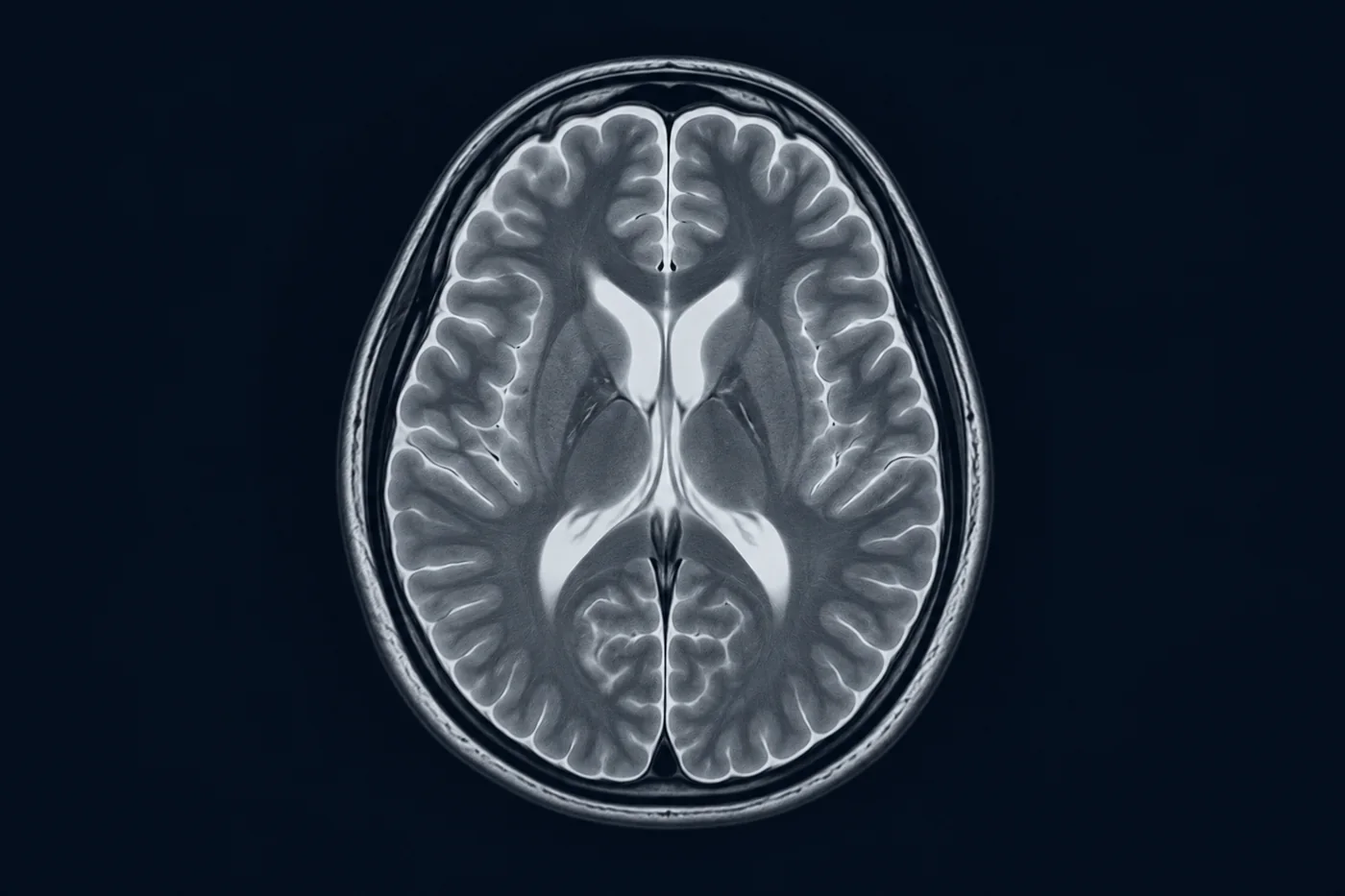
Brain MRI: the sequences that matter
Read T1, T2, FLAIR and diffusion with confidence.
Key points
- T1: fat is bright and CSF dark; best for anatomy and post-contrast images.
- T2: water and CSF are bright; most pathology is bright.
- FLAIR: T2 with CSF suppressed, so lesions next to the ventricles stand out.
- Diffusion-weighted imaging turns bright within minutes of ischaemic stroke.
- True restricted diffusion is bright on DWI and dark on the ADC map.
Image guide
What to look for in each view of this topic.
- T1-weighted. Grey matter darker than white matter, CSF black.
- T2-weighted. CSF bright; oedema and most lesions bright.
- FLAIR. Periventricular multiple sclerosis plaques perpendicular to the ventricles (Dawson's fingers).
- DWI and ADC. Acute left MCA infarct: bright on DWI, dark on ADC.
- Gadolinium T1. Ring-enhancing lesion: abscess or high-grade glioma; abscess restricts diffusion centrally.
- Susceptibility. Microbleeds and haemosiderin appear as dark blooming foci.
Related
Guideline summary8 min readAcute ischaemic stroke: reperfusion
Time is brain: image fast, thrombolyse eligible patients, and refer large-vessel occlusions for thrombectomy.
 Flashcards12 cards
Cranial nerves: function and lesions
One card per nerve: what it does and what a lesion looks like.
 Video12-min outline
Coronary circulation in 12 minutes
Map each coronary artery to the myocardium and ECG leads it supplies.