

Atlas · Respiratory · Image guide
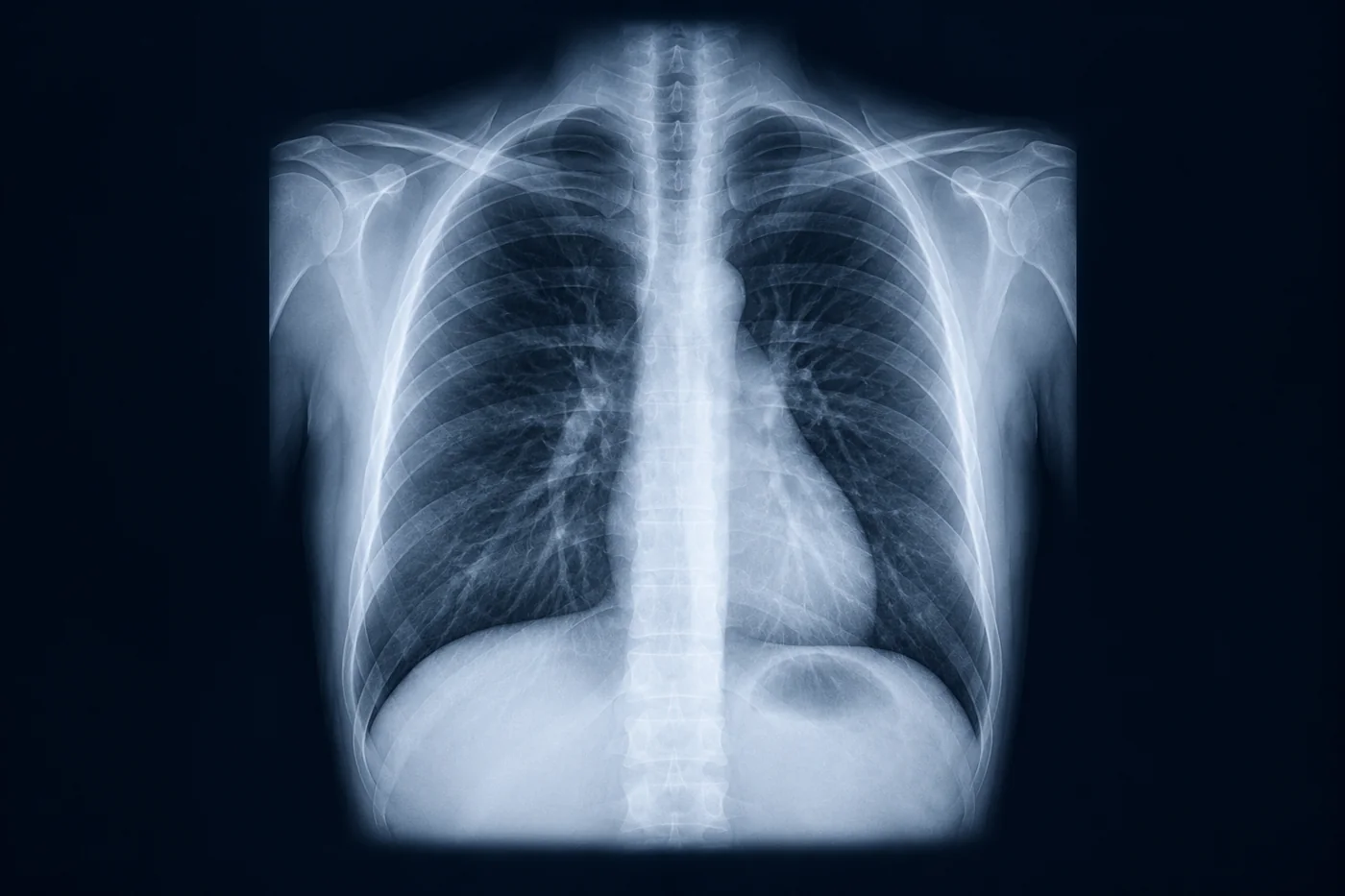
Chest X-ray: a systematic approach
A repeatable ABCDE read so you never miss the second finding.
Key points
- Check the patient details, date, projection (PA or AP), rotation, inspiration and penetration first.
- A: airway and trachea; the trachea deviates away from a tension pneumothorax and towards collapse.
- B: breathing; compare both lungs zone by zone, including behind the heart and below the diaphragm.
- C: circulation; a cardiothoracic ratio above 0.5 on a PA film suggests cardiomegaly.
- D and E: diaphragm (free air, blunted angles), then bones, soft tissues, lines and tubes.
Image guide
What to look for in each view of this topic.
- Normal PA film. Clavicle heads equidistant from the spinous processes (no rotation), 5–6 anterior ribs visible above the diaphragm.
- Right upper lobe collapse. Elevated horizontal fissure and tracheal shift to the right.
- Left lower lobe collapse. Triangular density behind the heart with loss of the medial left hemidiaphragm (sail sign).
- Tension pneumothorax. Hyperlucent hemithorax without lung markings, mediastinal shift away from the affected side.
- Pleural effusion. Meniscus at the costophrenic angle; about 200 mL is needed to blunt the angle on a PA film.
- Pneumoperitoneum. Crescent of free gas under the right hemidiaphragm on an erect film.
Related
 Guideline summary6 min read
Acute asthma in pregnancy
Treat the mother fully: it is the best treatment for the fetus.
Guideline summary8 min readPulmonary embolism: risk stratification
From pre-test probability to thrombolysis.
Notes7 min readSpirometry and lung function
Obstructive, restrictive or mixed — and what the transfer factor adds.