

Atlas · Gastrointestinal · Image guide
Liver histology essentials
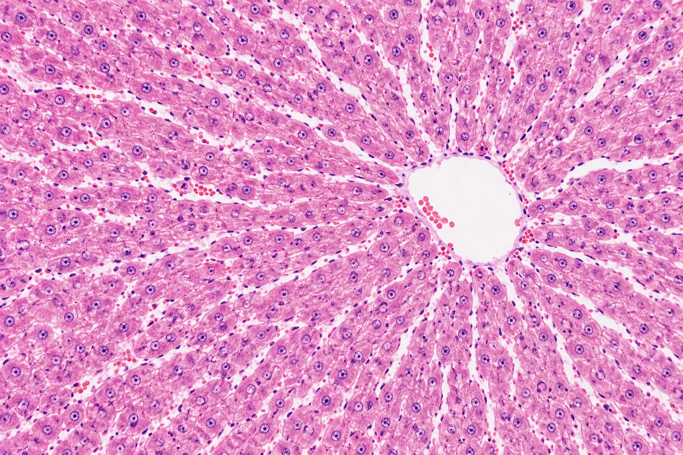
Lobules, acini and why the zones matter for injury patterns.
Key points
- Blood flows from the portal triad towards the central vein; bile flows the opposite way.
- Zone 1 (periportal) is oxygen-rich and affected first in viral hepatitis and by ingested toxins such as iron.
- Zone 3 (centrilobular) has the most cytochrome P450 and the least oxygen.
- Ischaemic hepatitis, paracetamol toxicity and alcohol injure zone 3 first.
- Kupffer cells are resident macrophages lining the sinusoids; stellate (Ito) cells store vitamin A and drive fibrosis.
Image guide
What to look for in each view of this topic.
- Portal triad. Branch of the hepatic artery, portal vein and bile duct in connective tissue.
- Central vein. Hepatocyte plates radiate towards the central vein, separated by sinusoids.
- Zone 3 necrosis. Centrilobular necrosis after paracetamol overdose or shock liver.
- Fatty change. Macrovesicular steatosis in alcohol-related and metabolic liver disease.
- Mallory–Denk bodies. Eosinophilic cytoplasmic inclusions of damaged keratin filaments in alcoholic hepatitis.
- Cirrhosis. Regenerative nodules surrounded by fibrous bands.
Related
Notes6 min readInterpreting liver tests
Hepatocellular or cholestatic, and what very high numbers mean.
 Video12-min outline
Coronary circulation in 12 minutes
Map each coronary artery to the myocardium and ECG leads it supplies.
 AtlasImage guide
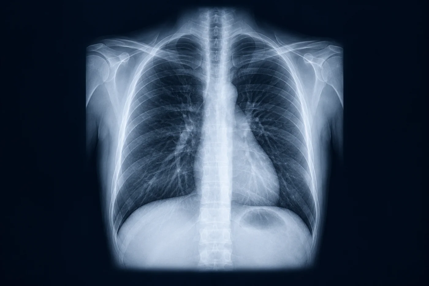
Chest X-ray: a systematic approach
A repeatable ABCDE read so you never miss the second finding.