

Atlas · Anatomy · 3D model
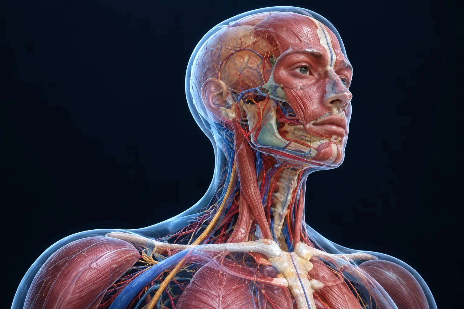
3D anatomy: muscles of the face and neck
Layer by layer, with nerve supply and clinical correlations.
Key points
- Muscles of facial expression are supplied by the facial nerve (CN VII).
- Muscles of mastication are supplied by the mandibular division of the trigeminal nerve (CN V3).
- Sternocleidomastoid and trapezius are supplied by the spinal accessory nerve (CN XI).
- Platysma is a facial-expression muscle in the neck, so it is supplied by CN VII.
- The carotid sheath contains the common (then internal) carotid artery, internal jugular vein and vagus nerve.
Image guide
What to look for in each view of this topic.
- Superficial layer. Platysma and muscles of facial expression from the second pharyngeal arch.
- Masticatory layer. Masseter, temporalis and the pterygoids from the first arch.
- Anterior triangle. Bounded by the midline, sternocleidomastoid and mandible; contains the carotid sheath.
- Posterior triangle. Contains the spinal accessory nerve, which is vulnerable in lymph node biopsy.
Related
 Video12-min outline
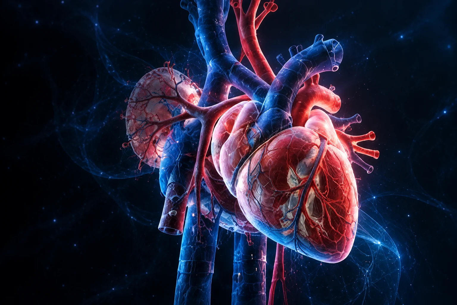
Coronary circulation in 12 minutes
Map each coronary artery to the myocardium and ECG leads it supplies.
 AtlasImage guide
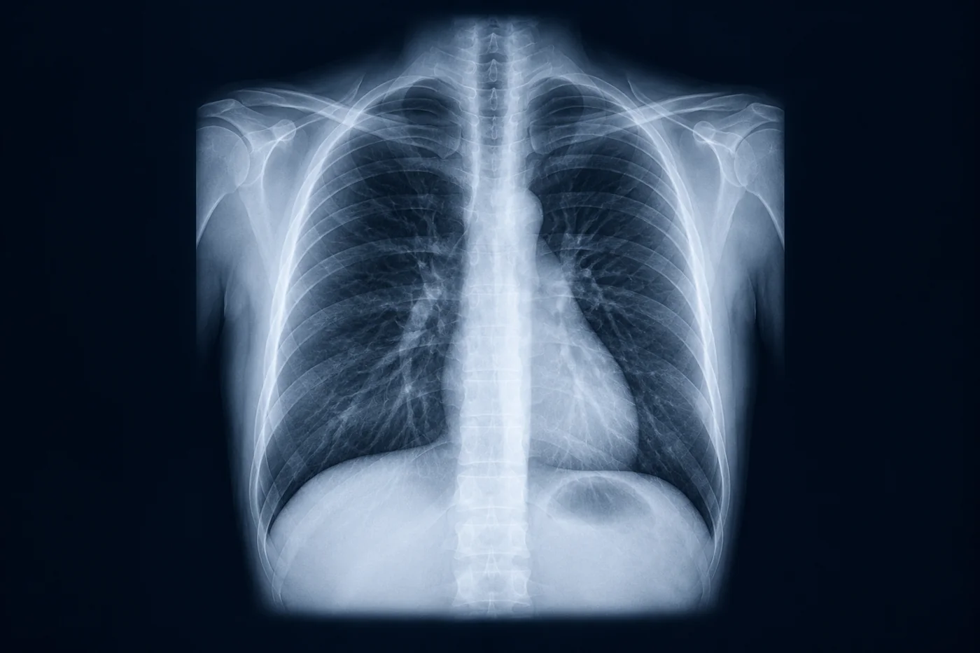
Chest X-ray: a systematic approach
A repeatable ABCDE read so you never miss the second finding.
 AtlasImage guide
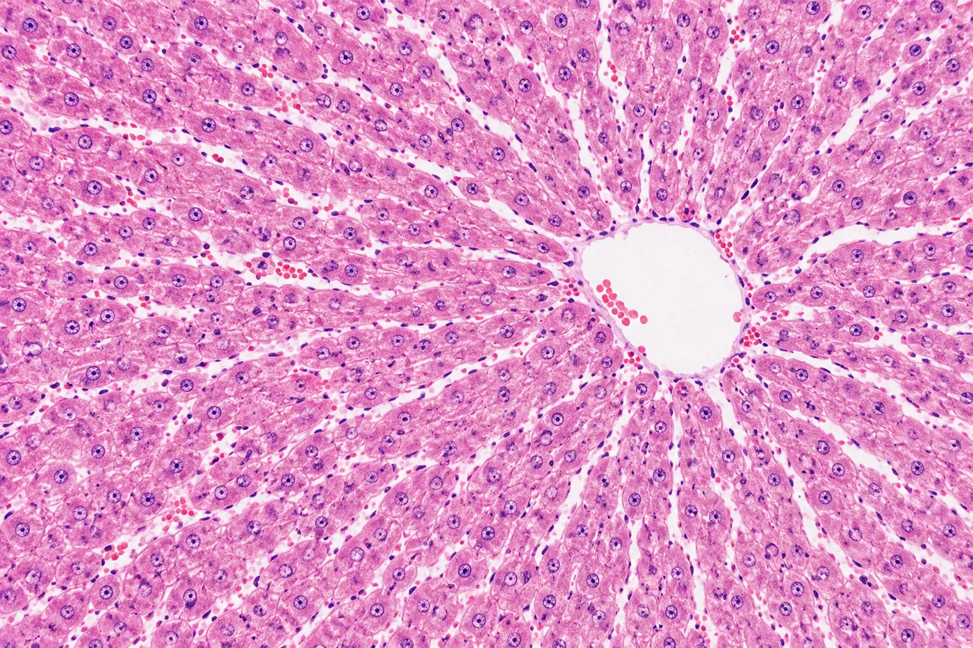
Liver histology essentials
Lobules, acini and why the zones matter for injury patterns.